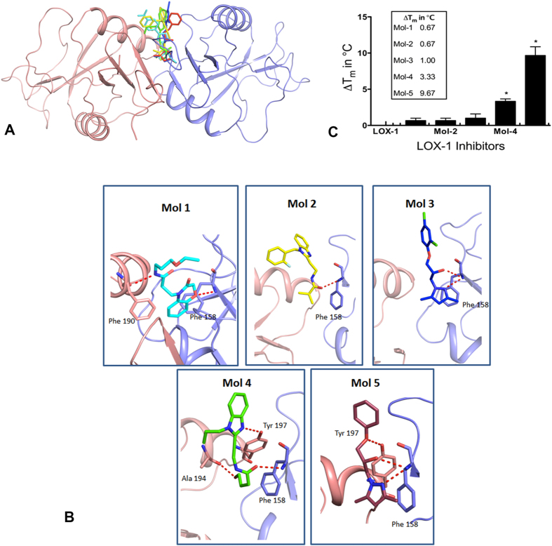
Figure 2. Docking of the 5 lead compounds and the thermal denaturation assay.
(A) The figure depicts the docking of the 5 lead compounds in the binding pocket of LOX-1. LOX-1 is shown as a ribbon model with one monomer in magenta and the other in blue. The compounds are shown as stick models in separate colors. (B) Hydrogen bonding interactions of the compounds as they dock to LOX-1. (C) The measured thermal shifts (in Celsius) are depicted for all five molecules. The solutions contained LOX-1 protein at 4 μM concentration. The ligand concentrations were 50 μM.

