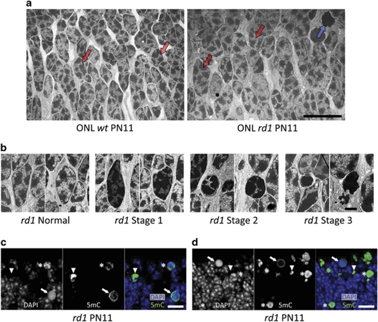
Figure 1.
Altered rd1 nuclear ultrastructure and DNA methylation. (a) The overview of PN11 wt and rd1 ONL illustrates the mixed distribution of heterochromatin and euchromatin (dark and light areas, respectively) in both wt and (most) rd1 photoreceptor nuclei. Typical examples of such nuclei are pointed out by red arrows. By contrast, rd1 photoreceptor nuclear configurations varied considerably from ‘normal' (=similar to wt nuclei), via the different stages 1 and 2, to very condensed, electron dense and dark, that is, stage 3 (blue arrow in rd1 picture). These stages are shown in more detail in panel (b). (c and d) In rd1 PN11 retina, immunostaining for 5mC (green), together with a nuclear counterstain (4,6-diamidino-2-phenylindole (DAPI), blue), showed varying degrees of co-localization. 5mC-positive structures had a DAPI appearance that was either heterogeneous rounded (*), or homogenous rounded (arrows), or weak to the point of being absent (arrowheads), most likely reflecting the different stages of nuclear condensation identified in panel (a). The confocal images in c and d are maximum projections of 16 and 21 Z-sections, respectively. Scale bars: a, c, and d=10 μm, b=2 μm

