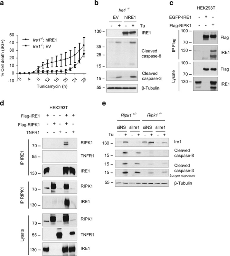
Figure 6.
RIPK1 interacts with the pro-apoptotic receptor IRE1. (a and b) Ire1−/− cells reconstituted with an empty vector (EV) or with a vector coding for hIRE1(hIRE1) were stimulated with 1 μg/ml tunicamycin (Tu), and the percentage of cell death was measured in function of time using the Fluostar Omega fluorescence plate reader (a), or cell lysates obtained after 17 h of stimulation were immunoblotted as indicated (b). (c) HEK293T cells were transiently transfected with plasmids coding for EGFP-hIRE1 and/or Flag-hRIPK1, and RIPK1 was immunprecipitated (IP) using anti-Flag-coated beads. Cell lysates and immunoprecipitates were analyzed by immunoblot as indicated. (d) HEK293T cells were transiently transfected with plasmids coding for Flag-hIRE1, Flag-hRIPK1 and/or hTNFR1, and IRE1 (upper panels) or RIPK1 (middle panels) were immunoprecipitated with anti-IRE1 or anti-RIPK1 antibodies, respectively. Cell lysates and immunoprecipitates were analyzed by immunoblot as indicated. (e) Ripk1+/+ and Ripk1−/− MEFs were transfected with a control non-silencing siRNA (siNS) or targeting Ire1 (siIre1) and then exposed to 1 μg/ml Tu for 12 h. The cells were then lysed and immunoblotted as indicated

