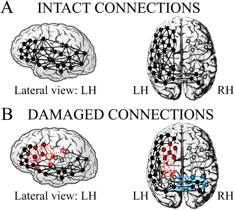
Figure 6.
Intra- and inter-hemispheric connections presumed/supposed to represent neural network model activated by linguistic circuits in (A) healthy controls and (B) non-fluent aphasic patients. Black nodes and connections: intact neural components; grey nodes and connections: (A) silent or less active centres in right hemisphere due to left-dominance for language and (B) inhibited/deafferented or less active centres in posterior left hemisphere due to the lesion on the main anterior linguistic centre. Red circles and dotted arrows (B): damaged non-functional areas. Cyan circles and arrows (B): re-activation or more active centres in right hemisphere as consequence of the lack of contralateral inhibition of posterior left centres (in grey) after deafferentation from damaged non-functional anterior areas (in red).

