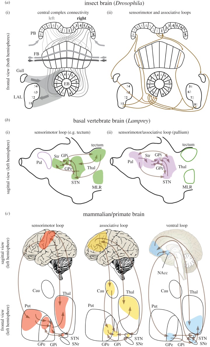
Figure 1.
Principal arrangements of the insect central complex, the lamprey and primate basal ganglia and their associated loops. (a) (i) Simplified schematic of the central complex (CX) showing connections between the protocerebral bridge (PB), fan-shaped body (FB) and ellipsoid body (EB), along with two satellite neuropils, the Gall and lateral accessory lobe (LAL). The noduli have been omitted. The PB is divided into synaptic modules which, depending on the species, vary between symmetrical arrangements of 9 + 9 (in Drosophila, Mantis religiosa) and as few as 5 + 5 (Notonecta) units. PB modules encode sensory representations (visual/tactile) from eight sectors on each side of the animal's long axis. Left and right representations of this ‘where’ code are relayed across eight modules of the FB such that left and right maps from the PB are compared. Outputs from the FB project into 16 modules of the EB such that modules representing opposite sectors are adjacent. The PB receives numerous inputs, including neurons entering laterally (arrows) carrying high-level information about visual motion direction. Columnar module in the FB are intersect by many dendritic trees and terminals (two shown) that likewise carry synthesized sensory information about complex parameters (‘what’ inputs) as well as a broad palette of modulatory peptides. The EB also receives a variety of inputs (also ‘what’ afferents), such as from the Gall and other satellite neuropils. Different combinations of where and what inputs result in different levels of activity in EB modules. Competing strengths of activity among EB modules result in a few achieving a stable output to the LAL. Further connections link the LAL system to pre-motor descending neurons (not shown). (ii) Comparison between sensorimotor and associative loops in insects (using the anatomy of the Drosophila as a general model) and sensorimotor, associative and ventral loops in mammals (using primates and humans in particular as a general model). Spatial organization is highlighted by the presence of numerically ordered modules in PB, FB, EB and LAL. The grey and bold black fonts indicate modules on the left and right side, respectively. (b) Anatomical representation of the re-entrant neural circuits characterizing sensorimotor and associative selections in the lamprey, which diverged already 560 Ma from the vertebrate lineages. (i) The first re-entrant neural circuit involves BG, thalamus and areas such as the optic tectum or the MLR, which provide sensorimotor inputs to the striatum via thalamus and receive direct inhibitory output from the BG. (ii) The circuit involves BG, thalamus and pallium, which projects directly towards the striatum and in turn receives mediated (via thalamus) inhibitory output from the BG. (c) Sensorimotor, associative and ventral (limbic) loops in mammals, here shown for primates. In the left hemisphere in humans, the different colours highlight the connectivity between separate areas in the cortex and their specific targets in the striatum and thalamus. This parallel partial segregation is maintained throughout the basal ganglia in the GP (globus pallidus), STN and SNr. Abbreviations: PB, protocerebral bridge; FB, fan shape body; EB, ellipsoid body; LAL, lateral accessory lobes; Pal, pallium; Str, striatum; MLR, mesencephalic locomotor region; Thal, thalamus; GPe, globus pallidus external segment, GPi, globus pallidus internal segment; STN, subthalamic nucleus, SNr, substantia nigra pars reticulata; Cau, caudate; NAcc, nucleus accumbens; Put, putamen.

