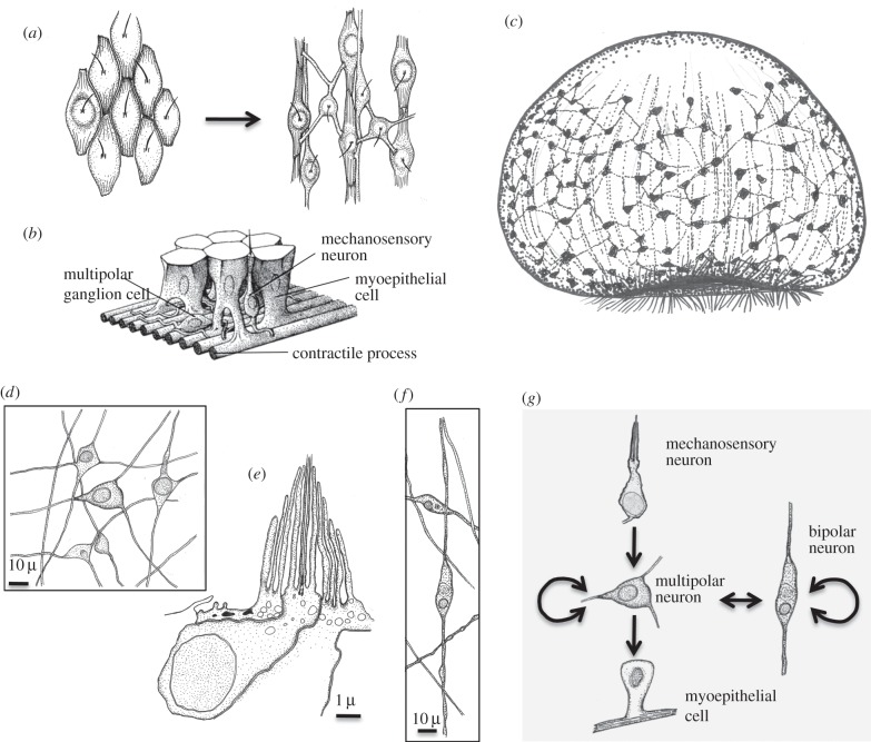
Figure 5.
Evolution of the nerve net. (a) Evolution of a sensory-contractile network of neurons and muscle cells by division of labour [5,6,125], following an initial scenario proposed by Mackie [126]. Starting point is a network of multifunctional, sensory-contractile cells in the gastraea ectoderm. Contractile fibres are oriented along the apical-blastoporal body axis. Individual sensory-motor neurons innervate many muscle cells. (b) Cell types of the cnidarian nerve net, modified from [127]. (c) The neuromuscular orthogon. Muscle cells have congregated into muscle strands. The evolution of true sensory, inter- and motorneurons enables the differential and antagonistic contraction of entire muscle strands as a response to remote stimuli. (d) Ganglion cells in the ectodermal nerve net in Cerianthus, after [128]. (e) Mechanosensory ciliary-cone receptors in the ectodermal nerve net in Cerianthus, after [56]. (f) Bipolar cells in the ectodermal nerve net in Cerianthus, after [128]. (g) Connections and signaling directions (arrows) in the nerve net of Cerianthus, after [128].

