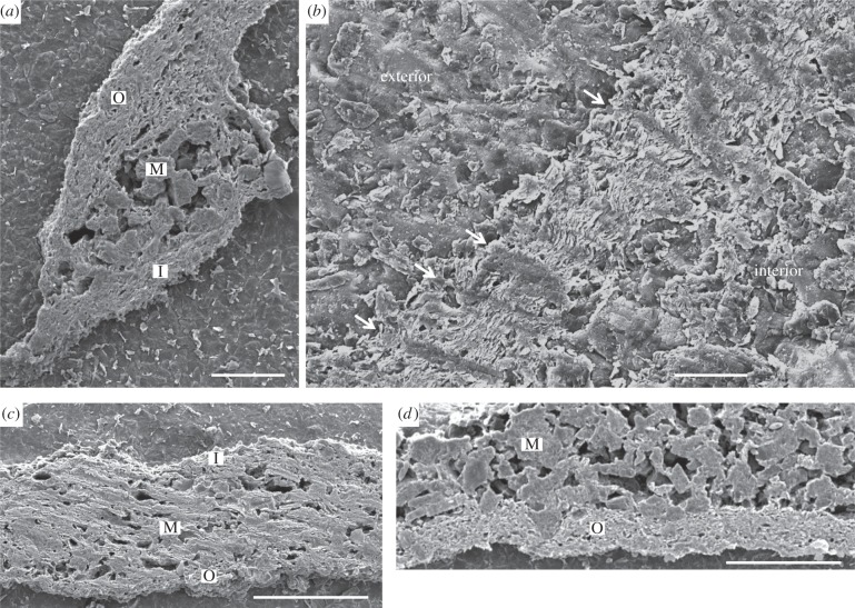
Figure 2.
Secondary emission SEM images of etched and polished transverse sections of Namacalathus hermanastes skeletal wall ultrastructure from the Nama Group, Namibia. (a,c,d), Tripartite organization. M, internal (middle) layer of rod-like microdolomite crystals; O, external outer foliated layers. I, inner foliated layers. (a) Scale bar, 200 µm. (c) Scale bar, 100 µm. (d) Scale bar, 100 µm. (b) Columnar microlamellar inflections (arrowed). Orientation with respect to the interior and exterior of the cup is noted. Scale bar, 200 µm.

