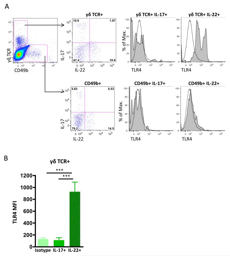
Figure 2. TLR4 expression by γδ T cells is associated with their production of IL-22 in mice with Collagen-Induced Arthritis.
Representative plots of the gating strategy for analysis of intracellular IL-17 and IL-22 expression by γδ TCR+ and CD49b+ NK T cells in the DLN from a single CIA mouse following ex vivo stimulation with PMA/ionomycin are shown (A). Expression of TLR4 was determined according to appropriate isotype controls (tinted grey histograms) in γδ T cells and CD49b+ NK T cells respectively. Data represent Mean Fluorescence Intensity (MFI) ± SD of DLN cells (B) from 5 individual mice (***p<0.001) undergoing CIA from a single experiment representative of two independent experiments

