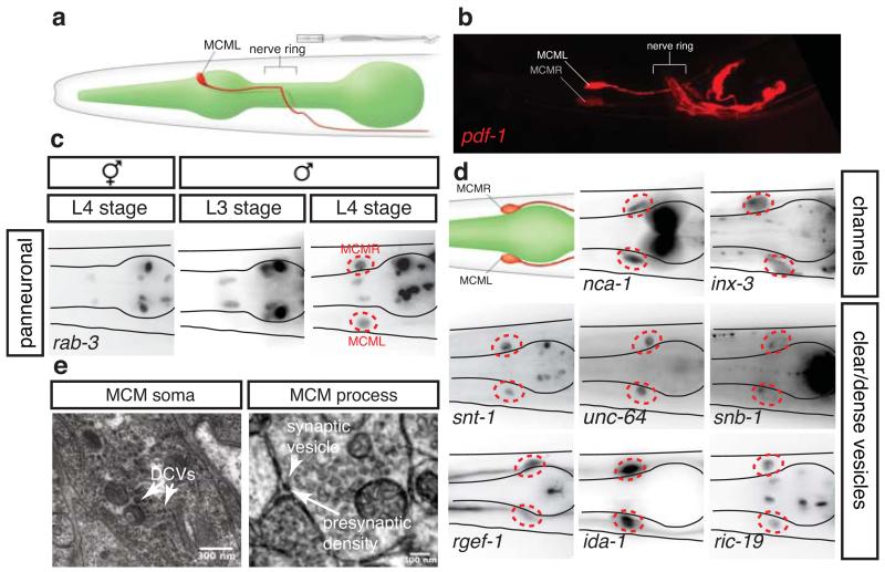
Figure 1. The MCMs are newly identified male-specific neurons.
Lateral views (a and b) and dorsal views (c and d) of animals oriented anterior to the left.
a, WormAtlas-style 11 diagram depicting the morphology and position, adjacent to the pharynx, of one of the bilateral pair of MCM neurons in the head of a male.
b, Confocal projection of pdf-1prom::rfp expression in the head of an adult male (same region as in a).
c, Expression of the pan-neuronal reporter transgene rab-3prom:rfp (Ras GTPase) in the head of an hermaphrodite and males at the third (L3) and fourth (L4) larval stages. The position of the MCMs is indicated with dashed red circles. R/L=Right/Left.
d, Expression in the MCMs in adult males of reporter transgenes for neuronal markers. snt-1 (synaptotagmin); unc-64 (syntaxin); snb-1 (synaptobrevin); rgef-1 (Ras exchange factor); nca-1 (NALCN Na+ channel subunit); inx-3 (gap junction innexin); ida-1 (tyrosine phosphatase-like receptor, phogrin); pdf-1 (neuropeptide pigment dispersing factor).
e, Electron micrographs of MCM showing dense-core vesicles (DCV) (left) and a synapse (right).

