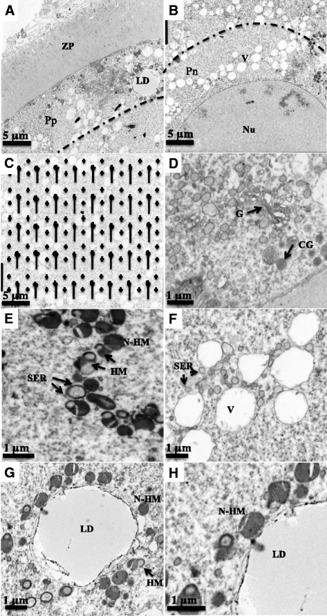
Fig. 2.

Electron micrographs (photographed at a primary magnification of 3000x) representing peripheral (Pp, a), perinuclear (Pn, b), and central (c) regions of ooplasm as well as the central region overlayed with the transparent grid for stereology (c). d, e, f, g, h illustrate cortical granules (CG), Golgi complex (G), hooded (HM) and non-hooded mitochondria (N-HM), vesicles (V), smooth endoplasmic reticulum (SER), and lipid droplets (LD) identified in the ooplasm for the quantitative data. Note the close spatial association between hooded-mitochondria (HM) and SER (e); and non-hooded mitochondria (N-HM) and lipid droplet (h). d, e, f and g represents peripheral ooplasm and (h) represent perinuclear ooplasm of an oocyte from D6W1 follicle, respectively. ZP = zona pellucida. Nu = nucleus
