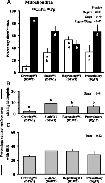
Fig. 3.

Mean ± SE of percent distribution of mitochondria in different regions of ooplasm (a), percentage of mitochondrial surface in contact with lipid droplets (b), and smooth endoplasmic reticulum (c) in oocytes collected at different stages of follicular growth and maturation: Day 3 of Wave 1 (D3W1), Day 6 of Wave 1 (D6W1), Day 1 of Wave 2 (D1W2) and preovulatory during estrus (Day ≥17). Pp and CnPn represent peripheral and central plus perinuclear regions of oocyte, respectively. abValues with no common superscript are different (P < 0.05)
