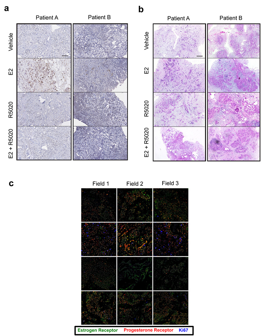
Extended data figure 8. Primary tumours cultivated as ex vivo explants shown response to progesterone.
Representative images of primary breast cancer explant tissue sections treated with vehicle, estrogen (E2), the progestin R5020 or estrogen plus progestin (E2 + R5020). These sections were probed with anti-Ki67 (brown) to label proliferating cells (a) or Haematoxylin and Eosin (b) . Each image is of a single tissue segment from a selection of 3-4 sections per sample treatment. Scale bar = 100mm. c. Confocal microscopy images (representative fields from each of the triplicate fragments) of a representative primary breast cancer explant tissue treated with vehicle, estrogen (E2), the progestin R5020 (Progestin) or estrogen plus progestin (E2 + R5020) and probed with anti-ERα (green), anti-PR (red) and anti-Ki67 to assess proliferating cells (blue).

