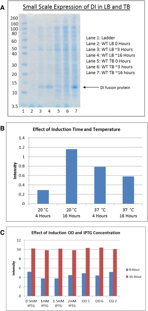
Fig. 2.

a SDS-PAGE gel comparing crude lysates of small scale expression of tagged Domain I fusion protein in LB and TB media before and after induction. Recombinant tagged DI fusion is indicated by an arrow and migrates at 12 kDa. Samples were lysed using triton/PBS and diluted in PBS (5x) and 20 μl was loaded onto an SDS PAGE Gel. The gel was then run, washed briefly in ddH2O and stained for 1 h with InstantBlue™ stain (Expedeon, UK). b & c Densitometric analysis was carried out on LB and TB samples on a small scale loaded in an identical way, gels were scanned, images converted to TIFF files and analysed. Values represent a calculation of intensity divided by area, OD denotes optical density of cultures at 600 nm at induction
