Fig. 3.

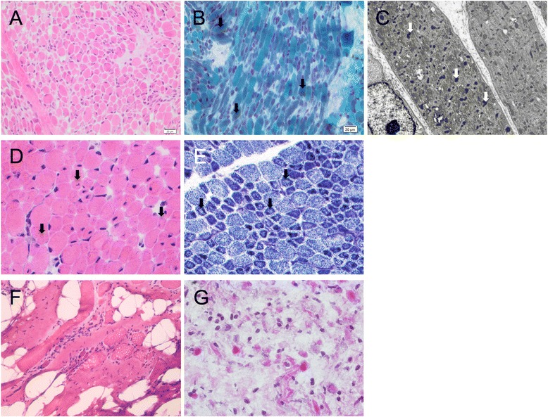
Histology of muscle biopsies from four families with mutations identified in the proband. Family 16 (a-c): h&e indicating variation in myofibre diameter (a) and Gomori trichrome staining showing dark purple regions suggesting nemaline bodies (arrows) (b). Electron micrograph, arrows indicate miliary nemaline bodies (c). (d) H&E stain of muscle from the proband in Family 14, indicating variation in myofibre size, central and internal nuclei. (e) Staining for NADH-TR in muscle from the proband in Family 14 with arrows indicating reduced central staining indicative of minicores. (f) H&E staining of muscle from the proband in Family 13 showing muscle tissue embedded in fibro-adipose tissue, with severe myopathic, non-specific changes. (g) H&E staining of muscle from the proband in Family 8, demonstrating a severe non-specific picture
