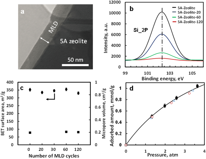
Figure 1. Characterization of 5A zeolite and 5A zeolite with molecular layer deposition (MLD) coatings.
(a) Transmission electron microscopy (TEM) image of 5A-Zeolite-60. (b) X-ray photoelectron spectra (XPS) of Si 2P of 5A zeolite and 5A zeolite with different cycles of MLD coating on 5A zeolite. (c) BET surface area of 5A zeolite and 5A zeolite with different cycles of MLD coatings (•), and micropore volume of 5A zeolite and 5A zeolite with different cycles of MLD coatings (■). Error bar is given automatically by Micromeritcs ASAP 2020 unit. (d) CH4 adsorption isotherms at 20 °C on 5A zeolite (■), 5A-Zeolite-30 ( ○), and 5A-Zeolite-60 (∆). Solid black line is a fit of adsorption points of CH4 on 5A zeolite by the Langmuir model. All MLD coatings have been calcined in air following the procedure described in the supplementary information.

