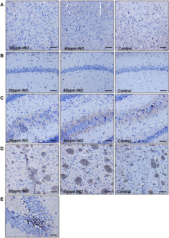
Fig. 6.

Representative photomicrograph of immunohistochemical staining for cleaved caspase 3 of the neocortex (a), hippocampal CA1 (b) and anterior and posterior CA 3/4 sectors (c), and basal ganglia (d). No caspase activation was found in any of the groups. Scale bar 90 μm. e Gyrus dentatus of an animal 6 hours after induction of subarachnoid hemorrhage as positive control for the staining. Scale bar 30 μm. Cerebral tissue harvested 7 days post cardiac arrest. iNO inhaled nitric oxide
