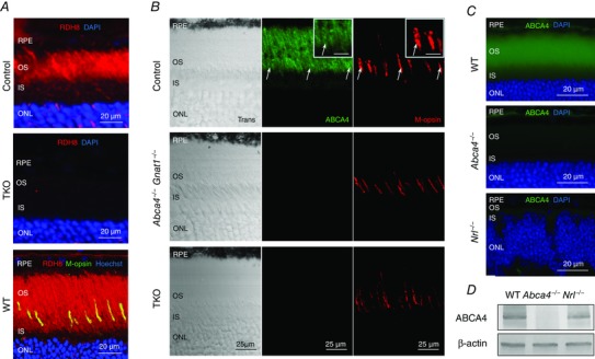
Figure 1. Expression of RDH8 and ABCA4 in rods and cones of 2‐month‐old mice .

A, RDH8 expression was examined by immunohistochemistry with anti‐RDH8 antibody. RDH8 (red) was present in Gnat1−/− (control) outer segments of photoreceptors, whereas no signal was detected in the retinas of Abca4−/−Rdh8−/−Gnat1−/− (TKO) mice. WT retinas revealed that mouse cones express RDH8 manifested as a yellow colour after co‐staining with anti‐RDH8 antibody (red) and anti‐M‐cone opsin antibody (M‐opsin, green). Cell nuclei (blue) were stained with DAPI or Hoechst 33342. Scale bar, 20 μm. B, immunostaining of control, Abca4−/−Gnat1−/− and TKO retinas (central location near the optic nerve) with Rim 3F4 anti‐ABCA4 antibody (green) or anti‐M‐opsin antibody (red). Scale bar, 25 μm. Trans, confocal images in transmitted light. Cell nuclei were stained with DAPI. White arrows indicate cone outer segments. Inset, higher magnification of cone outer segments. Scale bar, 10 μm. RPE, retinal pigmented epithelium; OS, outer segments; IS, inner segments; ONL, outer nuclear layer. C, immunostaining of WT, Abca4−/− and Nrl−/− retinas with TMR1 anti‐ABCA4 antibody (green). Scale bar, 20 μm. Cell nuclei were stained with DAPI. D, immunoblotting of WT, Abca4−/− and Nrl−/− retina samples using TMR1 anti‐ABCA4 antibody demonstrates that ABCA4 protein is expressed in Nrl−/− retinas. β‐actin served as a loading control.
