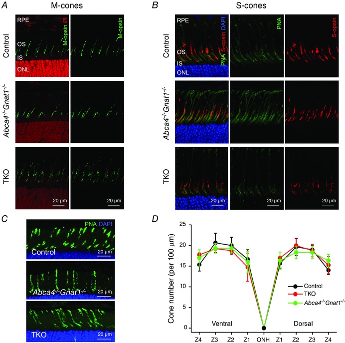
Figure 2. Normal cone density, morphology and opsin localization in 2‐month‐old mice lacking either ABCA4 or both ABCA4 and RDH8 .

Both M‐opsin (A), and S‐opsin (B) were properly localized in cone outer segments of Abca4−/−Gnat1−/− and TKO mice. Cross section images of central retina (near the optic nerve head) are shown. RPE, retinal pigmented epithelium; OS, outer segments; IS, inner segments; ONL, outer nuclear layer. Cell nuclei were stained with either propidium iodide (PI) or DAPI. Cone glycoprotein sheaths in B were additionally stained with PNA (green). Scale bar, 20 μm. C, cones in dorsal‐to‐ventral retina cross‐sections were stained with PNA (green) and nuclei were stained with DAPI (blue). Representative images from ventral retina (zone 2) are shown. Scale bar, 20 μm. D, PNA‐positive cells in 100 μm retina width were counted in four zones (Z1–Z4) of the dorsal and ventral retina areas. Z1, 400–500 μm; Z2, 900–1000 μm; Z3, 1400–1500 μm; and Z4, 1900–2000 μm from the optic nerve head (ONH). Error bars are SDs (control, n = 3; TKO, n = 4; Abca4−/−Gnat1−/−, n = 3).
