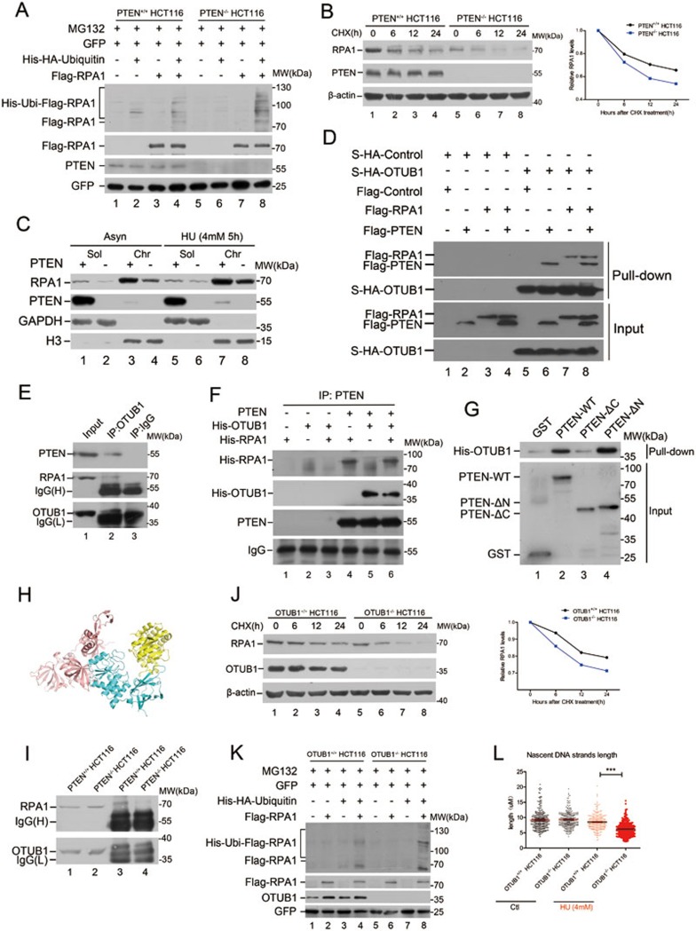
Figure 4.
PTEN promotes RPA1 protein stability by binding and recruiting OTUB1. (A) In vivo ubiquitination assay of RPA1. PTEN+/+ and PTEN−/− HCT116 cells were co-transfected with Flag-RPA1, His-HA-ubiquitin and GFP, pulled down with Ni-beads and immunoblotted with an antibody against Flag and other antibodies as indicated. Cells were treated with MG132 (10 μM) for 12 h before collection. (B) Half-life analysis of RPA1. PTEN+/+ and PTEN−/− HCT116 cells were treated with 100 μg/ml CHX, collected at different time points and immunoblotted with antibodies against RPA1, PTEN or β-actin. Graph shows quantification of RPA1 protein levels. RPA1 protein half-life was shortened in PTEN deficient cells. (C) Chromatin fraction analysis in PTEN+/+ and PTEN−/− HCT116 cells with or without HU treatment. Asynchronized (Asyn) or HU-treated PTEN+/+ and PTEN−/− HCT116 cells were subjected to fractionation. Soluble (sol) and chromatin (chr) fractions were separated and immunoblotted with indicated antibodies. (D) Exogenous binding of PTEN, RPA1 and OTUB1. S-tagged-OUTB1 co-transfected with Flag-RPA1, Flag-PTEN, or both, or Flag-mock was pulled-down with s-protein beads. A Flag specific antibody was used to detect exogenous RPA1 and PTEN. (E) Examination of physical interaction of PTEN, RPA1, and OTUB1. OTUB1 immunoprecipitates were subjected to western blotting using anti-PTEN and anti-RPA1 antibodies. (F) In vitro binding assay examining PTEN's interaction with His-tagged-RPA1 and His-tagged-OTUB1 and both. (G) In vitro binding assay with GST-tagged full-length PTEN, various GST-tagged PTEN domains and His-tagged-OTUB1. PTEN binds to OTUB1 with its C2 domain. (H) Illustration of in silico docking analysis of the PTEN/OTUB1/RPA complex to within a distance of 3.0 Å. PTEN is represented by cyan, RPA by salmon and OTUB1 by yellow. (I) In vivo binding of OTUB1 and RPA1 in PTEN+/+ and PTEN−/− HCT116 cells. OTUB1 immunoprecipitates in PTEN+/+ and PTEN−/− HCT116 cells were subjected to western blotting using an anti-RPA1 antibody. (J) Half-life analysis of RPA1 protein. OTUB1+/+ and OTUB1−/− HCT116 cells were treated with 100 μg/ml CHX, collected at different time points and immunoblotted with antibodies against RPA1, OTUB1, and β-actin. Graph shows quantification of RPA1 protein levels. RPA1 protein expression is decreased and its half-life shortened in OTUB1 null cells. (K) In vivo ubiquitination assay of RPA1. OTUB1+/+ and OTUB1−/− HCT116 cells were co-transfected with Flag-RPA1, His-HA-ubiquitin and GFP, pulled-down with Ni-beads and immunoblotted with antibody against Flag and other antibodies as indicated. Cells were treated with MG132 for 12 h before collection. (L) IdU tract lengths in OTUB1+/+ and OTUB1−/− HCT116 cells with or without HU treatment. IdU tracts measured in OTUB1−/− HCT116 cells show significant strand shortening with HU treatment as compared with those in OTUB1+/+ HCT116 cells. Data are presented as means ± SEM and analyzed by unpaired t-test. ***P< 0.001.

