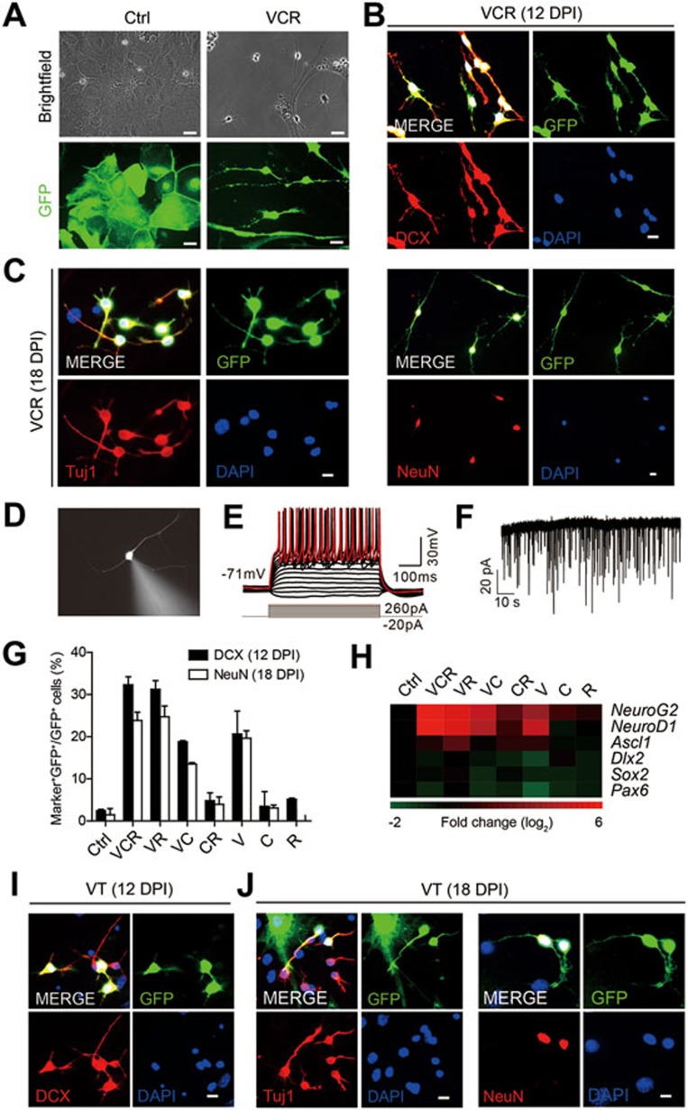
Figure 1.
Cultured astrocytes are induced into neuronal cells by small molecules. (A) Cultured astrocytes showed neuronal morphology 12 days post VCR induction (DPI, days post induction). GFP+ cells stand for astrocytes labelled by GFAP::GFP. (B) Cultured astrocytes were converted into DCX+ neuoroblasts 12 days post VCR induction. (C) Neuronal cells converted from astrocytes expressed mature neuron markers Tuj1 and NeuN 18 days post VCR treatment. (D) Patch-clamp recording was conducted on VCR-induced neurons from cultured astrocytes identified by fluorescence 26 days post induction. (E) Current-clamp recordings of neurons derived from cultured astrocytes with VCR treatment showed a representative train of action potentials with stepwise current injection. (F) Representative traces of spontaneous postsynaptic currents recorded in VCR-induced neurons from astrocytes. (G) Proportion of GFP+ cells co-expressing DCX or NeuN in the final GFP+ cells. (H) Heat map depicting the relative fold changes of gene expression levels in astrocytes 1 week after chemical treatment in vitro. The value in the color bar indicates log2 changes (relative to HPRT and normalized to ctrl). (I, J) DCX+, Tuj1+ and NeuN+ cells were generated from cultured astrocytes under treatment of the drug cocktail VT. Data are presented as mean ± SEM; scale bar, 10 μm.

