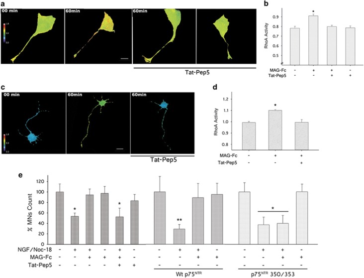
Figure 8.
MAG-Fc protects MNs from apoptosis via p75NTR-dependent activation of RhoA/ROCK signaling pathway. RhoA activity in MN1 cells and primary MN cultures transfected with a FRET-based RhoA biosensor. After 18 h, some cultures were pretreated for 60 min with 200 nM TAT-Pep5, a specific inhibitor of p75NTR–RhoA interaction. Then MNs were treated with 20 μg/ml of MAG-Fc for 1 h. (a) The images illustrate the pseudo-color thermal map of RhoA activation on MN1 cells (Scale bar, 22 μm). (b) Quantitative analysis of RhoA activity on MN1 cells. Data represent the mean±S.E.M. of three independent experiments (n=15 cells per group) (*P≤0.05). (c) Images depict the pseudo-color thermal map of RhoA activation on primary MN cultures (Scale bar, 20 μm). (d) Quantification of RhoA activity in MN cultures. Data represent the mean±S.E.M. of three independent experiments (n=12 cells per group; *P≤0.05). MN1 cells and primary MN cultures pretreated with TAT-Pep5 failed to show a MAG-Fc-dependent increase in RhoA activity. (e) Quantification of survival of MN1 cells pretreated with TAT-Pep5 or expressing Wt p75NTR or its double mutant form 350/353 and further treated with 20 μg/ml of MAG-Fc prior to the induction of apoptosis by treating cells with 100 nM NGF plus 50 nM NOC-18 for 24 h. (*P≤0.05; n=3)

