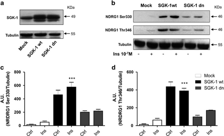
Figure 1.
Activity of SGK-1 in HEK-293 cells. Expression of SGK-1 protein is evaluated in HEK-293 cells stable transfected with pcDNA3 (Mock), SGK-1wt and SGK-1dn constructs. 100 μg of total extract protein was analysed by western blot using anti-SGK-1 antibody. Tubulin was used as a loading control (a). Activity of SGK-1 in HEK-293 cells stable transfected with pcDNA3 (Mock), SGK-1wt and SGK-1dn constructs was detected through NDRG1 phosphorylation levels in Thr346 and Ser330 in the presence and absence of Insulin 10−7 M for 30 min using western blot analysis. Tubulin was used as a loading control. (b) A quantification of three independent experiments by scanning densitometry is shown in (c and d), ***P<0.001 Ins (SGK-1 wt) versus Ctrl and Ins (Mock), and Ctrl and Ins (SGK-1 dn); N=3. Results are expressed as means±S.D.

