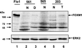
Fig. 1.

FOXM1 protein expression in gastric tissue. Western blots of FOXM1 protein expression in the indicated tissue type are shown. Molecular weights are in kDa. Results of normal gastric mucosa (N) and Tumour (T) or high grade dysplasia (D) in specimens 571,565,187 from three gastrectomies are shown. Flo1 cell lysate is shown in the left lane. ERK2 was used as a loading control
