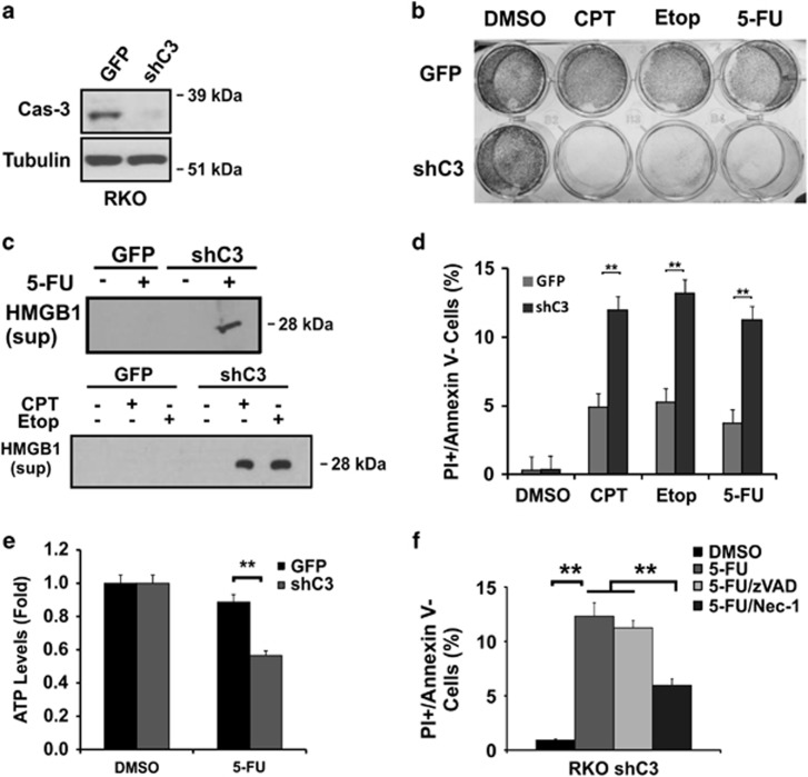
Figure 6.
RKO caspase-3 KD cells are sensitized to DNA-damaging agents. (a) Stable caspase-3 knockdown (shC3) RKO cells confirmed by western blotting. (b) Crystal violet staining of adherent RKO GFP or shC3 cells 48 h after CPT (500 nM), Etoposide (50 μM) or 5-FU (50 μg/ml) treatment. (c) Levels of HMGB1 in the medium from cells at 48 h with or without 5-FU treatment. (d) Fractions of PI+/Annexin V- cells as treated in (c). (e) ATP levels in cells as treated in (c). (f) Fractions of PI+/Annexin V- cells 48 h after 5-FU treatment, with or without z-VAD (20 μM) or Nec-1 (15 μM). (d–f) The data are mean + S.E.M. of triplicate wells. **P<0.01

