Figure 2.
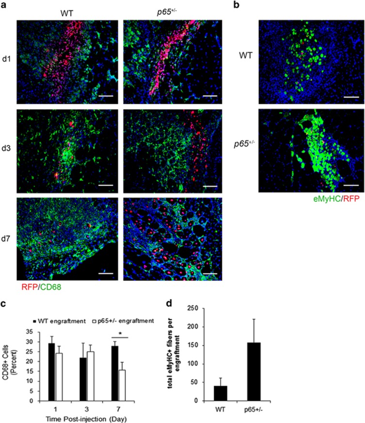
Donor p65+/− MDSC engraftments promote the repair of recipient muscle. (a) Immunofluorescent staining of tissue for the macrophage marker CD68 (green) indicated that RFP+ donor cell (red) engraftments in injured muscle were infiltrated by macrophages within 24 h postinjection (48 h postinjury), which continued to persist at 7 days (bottom). (b) eMyHC+ fibers (green) could be identified in or around donor cell engraftments at 7 days. (c) Quantification of CD68 positivity within x20 images indicated that p65+/− MDSC engraftments have significantly less CD68+ cells present at 7 days (*P⩽0.05), and (d) demonstrated a trend towards higher numbers of total eMyHC+ fibers (host+donor) (P=0.12). Data are displayed as mean±S.E.M.; n=3–4 mice per group. Scale bar: 100 μm

