Figure 5.
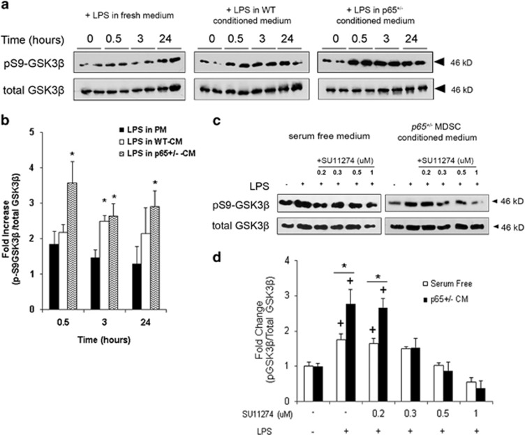
p65+/−-CM activates an HGF/Met/GSK3β pathway in RAW cells. (a) Western blot demonstrated that activation of RAW cells in PM induced an increase in pS9-GSK3β within 30 min (left), a response that was amplified by both WT- (middle) and p65+/−-CM (right). (b) Densitometric analysis revealed that when activated in p65+/−-CM, the fraction of pS9-GSK3β increased by 3.5-fold in 30 min, an amount significantly higher than WT-CM and PM groups (*P⩽0.05). (c) Inhibition of Met by SU11274 blocked pS9-GSK3β in RAW cells 30 min after exposure to LPS and p65+/−-CM (d) in a dose-dependent manner (*versus SF+LPS, +versus no LPS, P<0.05) Data are represented as mean±S.E.M. of at least three independent experiments

