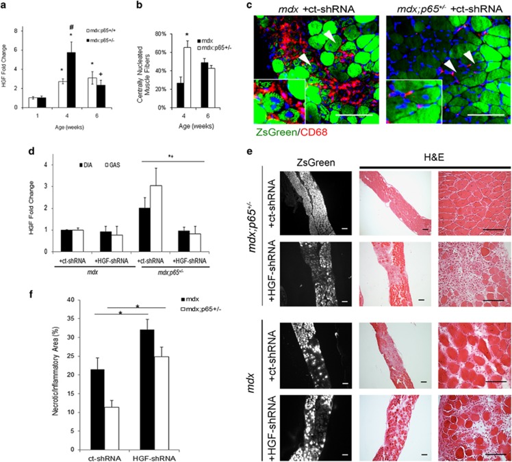
Figure 7.
Upregulation of Hgf in mdx;p65+/− skeletal muscle at 4 weeks of age correlates with accelerated regeneration. (a) Real-time RT-PCR demonstrated that Hgf expression was elevated in mdx:p65+/− muscle at 4 weeks, coinciding with enhanced regeneration, quantified as the percent of centrally nucleated muscle fibers in (b) (*versus WT, P⩽0.05; +versus WT, P⩽0.10; #versus mdx, P⩽0.05). (c) ZsGreen+ centrally nucleated fibers indicated that muscle progenitor cells were transduced by the AAV vector (arrows, top). (d) After 4 weeks, Hgf expression was significantly reduced in the DIA and GAS muscles of HGF-shRNA-treated mdx;p65+/− mice compared with the ct-shRNA-treated group (P⩽0.05, +shHGF versus ct-shRNA; *GAS, +DIA). (e) Silencing of Hgf worsens the histopathology of the DIA muscle from treated mdx;p65+/− mice (top) and mdx mice. (f) We quantified the necrotic/inflammatory lesions in H&E-stained DIA tissue sections from treated mice by measuring the lesions as percent area; n=4–6 mice per group. Data are displayed as mean±S.E.M.

