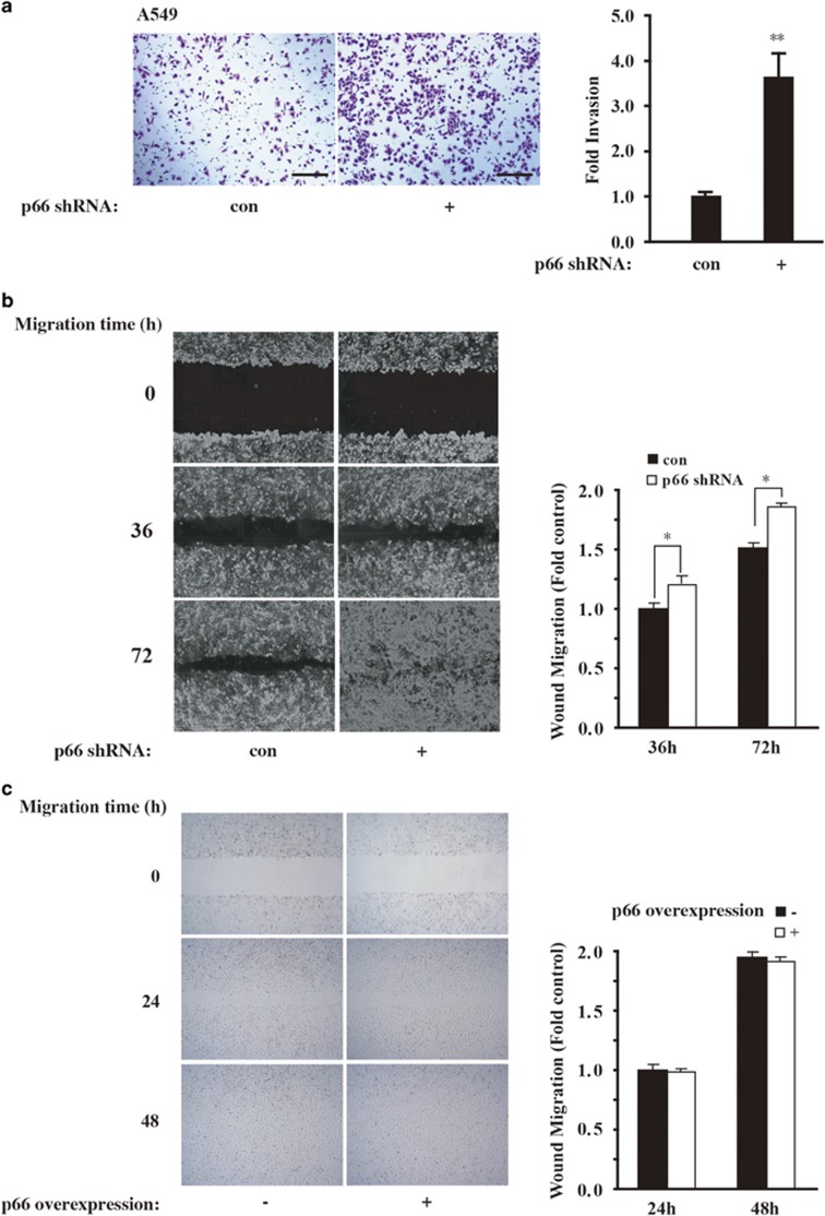
Figure 4.
p66Shc depletion increases cell invasion and migration. (a) Boyden chamber assay for A549 cells transduced with control or p66Shc shRNA as described in Materials and methods. Scale bars, 100 μm. Quantification of invasion change are shown in the right panel. **P<0.01 as compared with the cells treated with control shRNA alone. (b) A549 cells were transduced as in a and plated on fibronectin-coated coverslips. Phase-contrast images were obtained immediately after wounding and at 36 and 72 h after wounding. Quantification of mean speed of cells migrating into the wound shown in the right panel. Error bars represent S.E.M. *P<0.05 as compared with the cells treated with control shRNA alone, based on three independent experiments. (c) A549 p66Shc-overexpressing cells were plated on fibronectin-coated coverslips. Phase-contrast images were obtained immediately after wounding and at 24 and 48 h after wounding as in b, respectively

