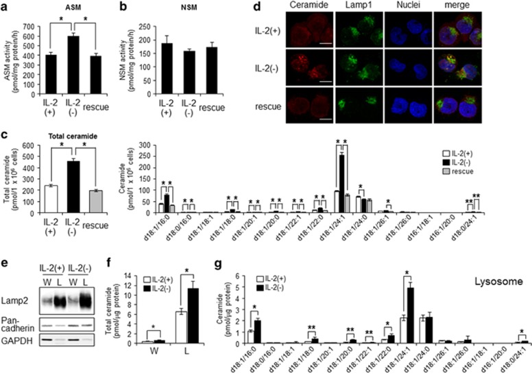
Figure 2.
IL-2(−) induced accumulation of lysosomal ceramide. (a and b) KHYG-1 cells were cultured in IL-2(+), IL-2(−), and rescue conditions for 12 h. Cells were harvested, and after extraction of the proteins, the activities of each enzyme were assessed. Activities of ASM (a) and NSM (b) were measured using C6-NBD-ceramide and C6-NBD-SM as substrates. (c) After 36 h, ceramide levels were assessed by LC-MS/MS. (d) Cells were cultured at each condition for 12 h, fixed, stained with anti-ceramide and Lamp1 (lysosome) antibodies, and analyzed by confocal microscopy. Nuclei were counter-stained with DAPI. Scale bars, 10 μm. (e) After 24 h, lysosome fraction were isolated as described in Materials and Methods section. Lamp2 (lysosome), pan-cadherin (plasma membrane), and GAPDH (cytosol) were used as markers for each organelle in whole cell lysate (W) and lysosome fraction (L). (f and g) Total ceramide levels in whole cell and lysosome (f) and ceramide species in lysosome (g) were assessed by LC-MS/MS. Values are mean±S.D. from three different experiments. *P<0.005, **P<0.05

