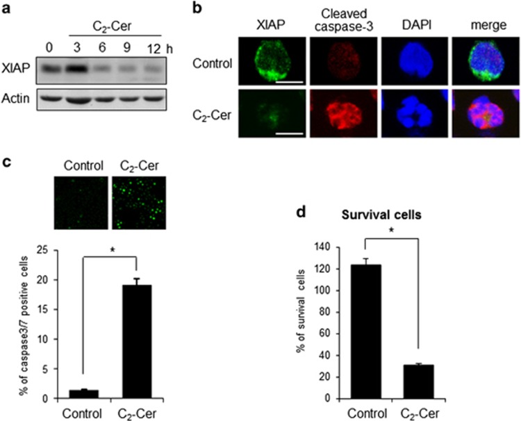
Figure 8.
C2-ceramide induced apoptosis with XIAP degradation. (a) KHYG-1 cells were treated with 50 μM C2-ceramide (C2-Cer) for the indicated times. XIAP protein was detected by western blotting analysis. (b) After 12 h of C2-Cer treatment, cells were stained with anti-XIAP and anticleaved caspase-3 antibodies. Nuclei were counterstained with DAPI. Images were obtained by confocal microscopy. Scale bars, 10 μm. (c) In vivo caspase-3/-7 activity was measured by fluorescent substrate of caspase-3/7 (FAM-DEVD-FMK). Images were obtained by fluorescent microscopy. Values are mean±S.D. from three different experiments. *P<0.005. (d) After 12 h, viable cell numbers were counted after staining with trypan blue and indicated as a percentage of vehicle treatment. Values were mean±S.D. from three different experiments. *P<0.005

