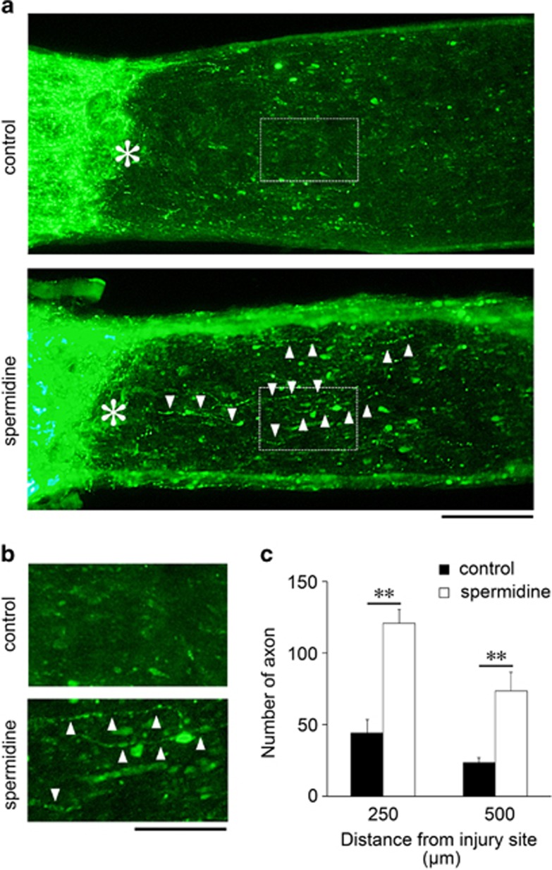
Figure 6.
Effects of spermidine on optic nerve regeneration following ONI in vivo. (a) Longitudinal sections through the optic nerve showing CTB-positive axons distal to the injury site (*) in control and spermidine-treated mice at d14 after ONI. Arrowheads indicate regenerating axons. Scale bar: 100 μm. (b) Higher magnification of the images shown in a. Scale bar, 50 μm. (c) Quantitative analyses of regenerating axons extending 250 and 500 μm from the injury site at d14 after ONI. The data are presented as means±S.E.M. of six samples for each experiment. **P<0.01

