Figure 1.
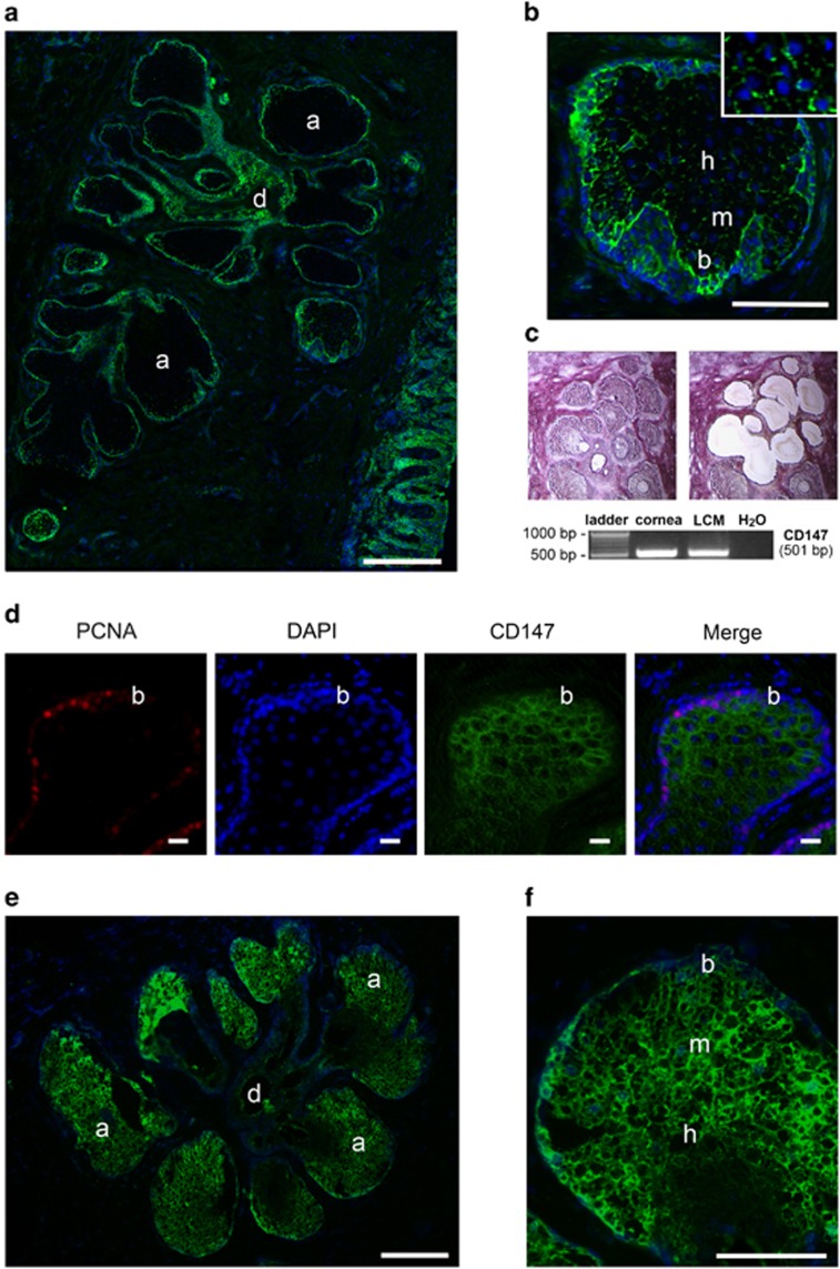
CD147 and gelatinolytic activity localize to acinar structures in human meibomian glands. (a) Immunofluorescence microscopy of a human eyelid showing CD147 localization (green) to basal cells at the peripheral margin of secretory acini and to the multilayered squamous epithelium of the ductule. Nuclei were counterstained using DAPI (blue). (b) Within the acinus, CD147 staining decreased towards differentiated mature and hypermature meibocytes. This central region was characterized by patchy staining of CD147 along the cell membrane (inset). (c) Laser capture microdissection (LCM) and RT-PCR analysis demonstrating CD147 mRNA in acinar structures of meibomian gland tissue. Representative micrographs are shown before (left) and after (right) microdissection. Cultures of human corneal epithelial cells were used as positive control. (d) Immunofluorescence microscopy of a human eyelid showing PCNA (red) localization to basal acinar cells. CD147 was labeled in green. Nuclei were counterstained using DAPI (blue). (e) In situ gelatin zymography showing localization of gelatinolytic activity in secretory acinar cells of the meibomian gland. (f) Within the acinus, activity was detected in basal and mature meibocytes, and decreased towards the disintegrating zone. Acinus (a), basal cell (b), ductile (d), epidermis (e), mature cell (m), hypermature cell (h). Scale bars, 300 μm (a), 10 μm (d), 200 μm (e), 100 μm (b and f)

