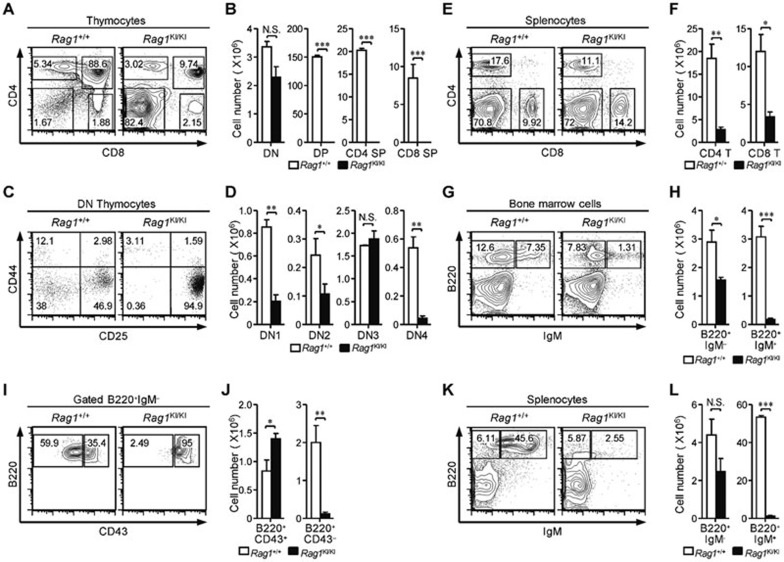
Figure 1.
Impaired T and B lymphocyte development in RAG1KI/KI mice. (A, B) Thymocyte development is arrested at the DN stage. Flow cytometric analysis of thymocytes, from the indicated mice, stained with anti-CD4 and anti-CD8 antibodies. The cell number of indicated thymocyte subsets is shown (mean ± SEM; n = 3; ***P < 0.001 by Student's t-test; N.S., no significance). (C, D) Thymocytes are primarily blocked at DN3 stage. Lin− cells were gated and analyzed for CD44 and CD25 expression. The cell number of the DN subsets is shown (mean ± SEM; n = 3; **P < 0.01 and *P < 0.05 by Student's t-test; N.S., no significance). (E, F) Mature T lymphocytes are reduced in the periphery. Splenocytes were stained with anti-CD4 and anti-CD8 antibodies. The number of CD4 and CD8 lymphocytes in spleen is shown (mean ± SEM; n = 3; **P < 0.01 and *P < 0.05 by Student's t-test). (G, H) Early B cell development is impaired. Cells isolated from femurs were stained with anti-B220 and anti-IgM antibodies. The number of indicated B cell subsets in the bone marrow is shown (mean ± SEM; n = 4; ***P < 0.001 and *P < 0.05 by Student's t-test). (I, J) B220+IgM− cells were gated and analyzed for CD43 expression. The number of pro-B-cell and pre-B-cell subsets are shown (mean ± SEM; n = 4; **P < 0.01 and *P < 0.05 by Student's t-test). (K, L) Mature B cells were barely detected in the spleen. Splenocytes were stained with anti-B220 and anti-IgM antibodies. The number of indicated B-cell subsets in spleen is shown (mean ± SEM; n = 4; ***P < 0.001 by Student's t-test; N.S., no significance). All mice were analyzed at 4-5 wk of age.

