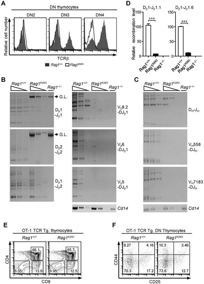
Figure 2.
Impaired Tcrb and Igh rearrangement in RAG1KI/KI mice. (A) The TCRβ expression level dropped markedly. Overlay of the TCRβ intracellular expression in the indicated DN subsets (solid line, RAG1KI/KI mice; shaded in gray, WT littermates). The results are representative of three independent experiments. (B) Tcrb rearrangement is reduced. Semi-quantitative PCR to detect Dβ-Jβ rearrangement or Vβ-DJβ1 recombination in DN3 thymocytes (CD4−CD8−CD44−CD25+) sorted from RAG1KI/KI mice, their WT littermates and RAG1−/− mice. Below, amplification of a Cd14 fragment (input control). G.L., germline fragment. PCR amplification was performed on serial fivefold dilutions of genomic DNA. The results are representative of three independent experiments. (C) Igh rearrangement is impaired. Semi-quantitative PCR of DH-JH rearrangement or VH-DJH recombination in Pro-B cells (B220+IgM−CD43high) sorted from the indicated mice. Below, amplification of a Cd14 fragment (input control). PCR amplification was performed on serial five-fold dilutions of genomic DNA. The results are representative of three independent experiments. (D) Quantification of the rearrangement products. Real-time PCR analysis quantification of the Dβ1-Jβ1.1 signal joints and Dβ1-Jβ1.6 coding joints (mean ± SEM; n = 3; ***P < 0.001 by Student's t-test). (E) Flow cytometric analysis of thymocytes from the indicated mice stained with anti-CD4 and anti-CD8 antibodies. The results are representative of two independent experiments. (F) CD4−CD8− cells were gated and analyzed for CD44 and CD25 expression. The results are representative of two independent experiments. All recombination products amplified by PCR were confirmed by sequencing.

