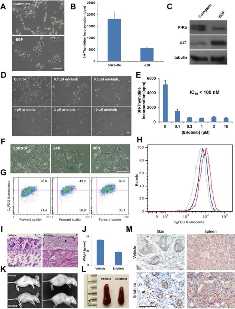
Figure 1.
Anti-senescence function of EGF in human epithelial cells and mouse tissues. (A-C) EGF depletion results in cellular senescence of HME cells. HME cells were exposed for 1 week to complete culture medium or to medium lacking EGF. Cells cultured in the absence of EGF displayed an enlarged morphology, elevated SA-β-gal activity (A), decreased proliferation (B), and reduced Rb phosphorylation and elevated p21 expression (C), all markers that are commonly associated with cellular senescence. (D-H) EGFR pharmacological inhibition causes cellular senescence of HME cells. In D and E, HME cells were treated for three days with complete culture medium supplemented with increasing concentrations of erlotinib. Cells were then stained for SA-β-gal activity (D), or metabolically labeled with 2 μCi of [3H]-thymidine for 16 h (E). Progressively decreasing amounts of label incorporation were observed for cells cultured in the presence of erlotinib. Experiments were performed in triplicate; error bars indicate SD. In F-H, quantification of erlotinib-induced cellular senescence of HME cells using flow cytometry. HME cells were exposed to 1 μM erlotinib for 24 or 48 h and then analyzed for SA-β-gal activity using either a chromogenic assay (F) or a fluorescence-based assay (G and H, see Supplementary information, Data S1). Both assay types indicated that SA-β-gal reached maximal activity from 24 to 48 h after treatment. (A, D, F) Scale bar, 50 μm. (I-M) Mice treated with erlotinib display early aging-associated phenotypes. Skin atrophy in erlotinib-treated mice. BALB/c nude mice (five per group) were treated with either vehicle or erlotinib (50 mg/kg/day) for 4 weeks. Skin was fixed in 4% PFA, stained using H&E, and photographed at 10× magnification. Note the reduced epidermal thickness and fat content resulting from EGFR inhibition. E, epidermis; D, dermis; F, fat; M, muscle. Scale bar, 250 μm. (I). Reduced weight of mice treated with daily erlotinib. Error bars indicate SD (J). Representative photograph and X-ray of tumor-bearing BALB/c nude mice treated with vehicle or erlotinib. Note the reduced size, kyphosis, and generally aged appearance of the erlotinib-treated mouse (K). Spleen atrophy in erlotinib-treated mice (see also Supplementary information, Figure S1P-S1Q) (L). Immunohistochemical staining of skin and spleen tissue from vehicle- or erlotinib-treated mice was performed using an antibody specific for p16. Images shown were photographed at 40×. Scale bar, 100 μm (M).

