Figure 1.
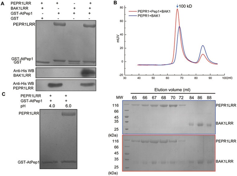
PEPR1 recognition of AtPep1 induces ectodomain heterodimerization of PEPR1 and BAK1. (A) In vitro reconstitution of AtPep1-induced PEPR1LRR-BAK1LRR heterodimerization using purified proteins. His-tagged PEPR1LRR and BAK1LRR proteins were expressed in insect cells and purified to homogeneity. The GST-AtPep1 was expressed in E. Coli. An equal amount of the purified GST- AtPep1 protein was loaded onto 30 μl of GS4B-resin and then incubated with an excessive amount of His-tagged PEPR1LRR and BAK1LRR on ice for 20 min. After extensive washing, proteins bound to the GS4B resin were detected by Coomassie blue staining or western blot. WB: western blot. GST in line 3 likely resulted from proteolytic removal of AtPep1. (B) AtPep1 induces PEPR1LRR-BAK1LRR heterodimerization in solution. Top panel: superposition of the gel filtration chromatograms of PEPR1LRR and BAK1LRR in the absence (blue) or presence (red) of AtPep1. The vertical and horizontal axes represent ultraviolet absorbance (λ = 280 nm) and elution volume (ml), respectively. The molecular weight for the PEPR1LRR-AtPep1-BAK1LRR complex is indicated. Bottom panels: Coomassie blue staining of the peak fractions shown on the top following SDS-PAGE. (C) AtPep1-PEPR1LRR interaction is pH-dependent. The assay was performed as described in A.

