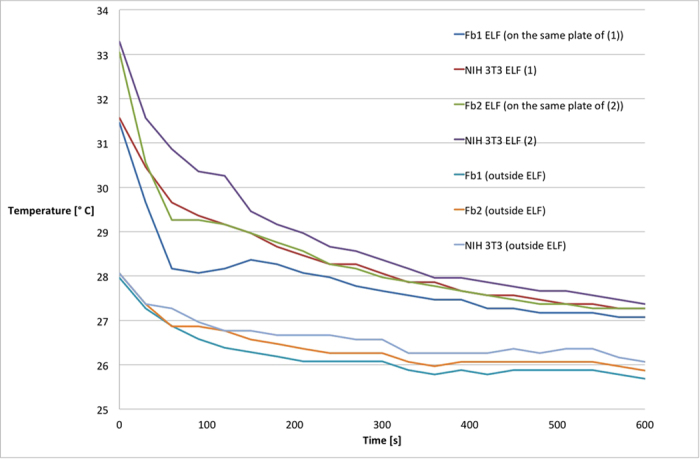
Figure 1. Thermal dispersion of cellular models.

Thermal dispersion was evaluated at room temperature by the infrared thermographic camera. Two different preparations of primary fibroblasts (Fb1 and Fb2) and NIH 3T3 cells were analysed in the same conditions, either exposed for 6 days to electromagnetic field (ELF) or not exposed. The figure is representative of a set of three independent experiments.
