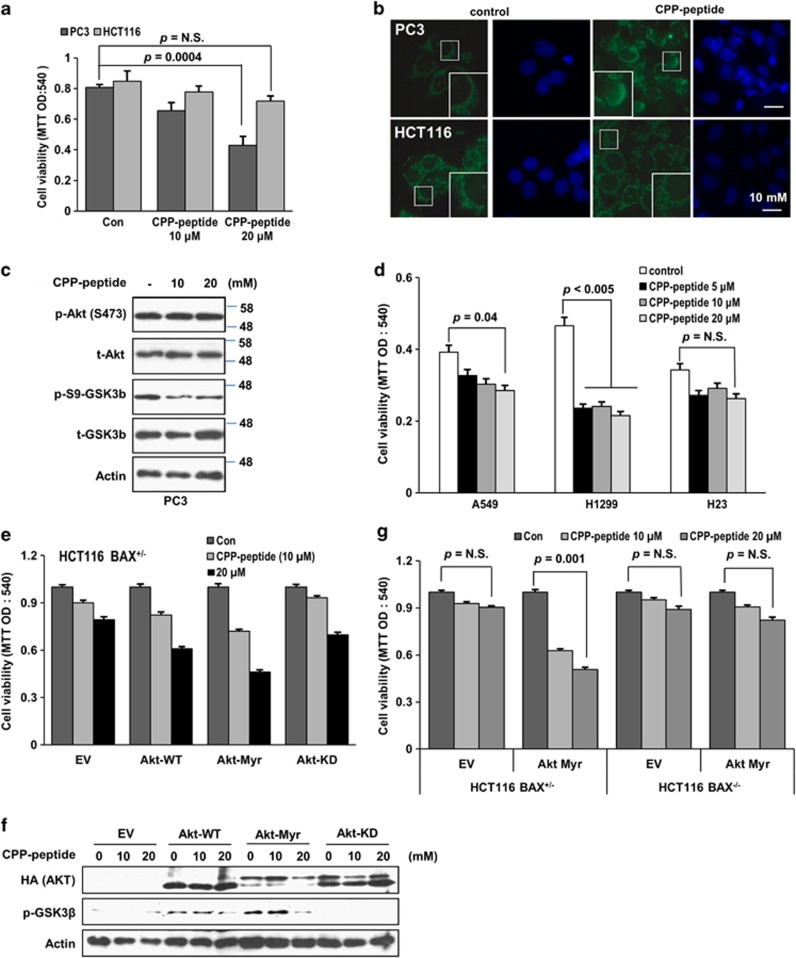
Figure 6.
CPP-fused BH3BIM(I155R/E158S) peptide induces death of PC3 cells. (a) Cell viability evaluated by the MTT assay 72 h after treatment of the peptide (0, 10 or 20 μM). N.S indicates non-significance. Error bars indicate S.D. (n=3). (b) The immunofluorescence images of PC3 and HCT116 cells treated with 20-μM CPP-BH3BIM(I155R/E158S). Cytochrome c (green) was detected by anti-cytochrome c antibody and FITC-conjugated secondary antibody, and the nucleus was stained by DAPI. The large boxes are the enlargement of the small boxes, highlighting the cytochrome c staining. (c) Western blot (WB) analysis to examine the effect of CPP-BH3BIM(I155R/E158S) on the phosphorylation of Akt (at Ser473) and GSK3β (at Ser9). PC3 cells were incubated with the peptide for 6 h at the indicated concentration. t-Akt and t-GSK3β stand for total Akt and GSK3β, respectively. (d) MTT assay to examine the cytotoxic effect of CPP-BH3BIM(I155R/E158S) on lung cancer cell lines. The viability of PTEN-silenced H1299 cells was clearly reduced by the peptide. (e) Promotion of CPP-BH3BIM(I155R/E158S)-induced death of HCT116 cells by Akt. Cells were transfected with the indicated vectors for 24 h and incubated with the peptide for 72 h. Cell death was promoted by Akt-WT and Akt-Myr, but not by Akt-KD. (f) Phosphorylation of GSK3β (p-GSK3β) by Akt-WT and Akt-Myr, but not by Akt-KD. Cells were harvested for WB after incubation with CPP-peptide for 6 h. Actin was used for loading control. (g) CPP-BH3BIM(I155R/E158S) induced apoptosis in a BAX-dependent manner. HCT116 cells and its isogenic BAX−/− cells were transfected with Akt-Myr for 24 h and incubated with the peptide at the indicated concentration for 72 h. HCT116 BAX+/− cells, but not HCT116 BAX−/− cells, were affected by the peptide

