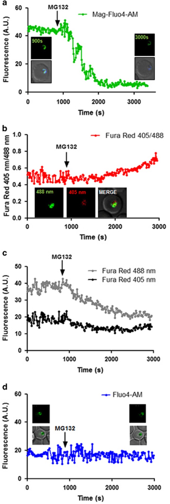
Figure 4.
ER stress-associated Ca2+ kinetics by confocal fluorescence microscopy. (a) Kinetics of free calcium release from the ER upon MG132 treatment as assessed by Mag-Fluo-4 AM staining. Parasites at early trophozoite stage were loaded with Mag-Fluo-4 AM and fluorescence intensity was plotted as a function of time. Treatment with MG132 (100 nM) resulted in a rapid loss of ER-free calcium. The experiments was repeated for 20 iRBCs in two independent experiments; a single selected representative graph and image (inset showing colocalization with ER Tracker Blue white) is shown. (b) Confocal live cell imaging with Fura-Red AM staining shows that cytoplasmic-free Ca levels rise after MG132 treatment (100 nM). A representative parasite's graph and image is presented here. The fluorescence ratio (405/488 nm) increases following MG132 addition. (c) Graph showing decrease in Fura-Red fluorescence at 405 and 488 nm for the above parasite. (d) Fluo-4 AM staining to demonstrate that there is no decrease in digestive vacuole calcium concentration during this period of MG132 treatment. The fluorescence intensity before and after addition of MG132 remains nearly constant. Additional fluorescence graphs and images are included in Supplementary Figure 2

