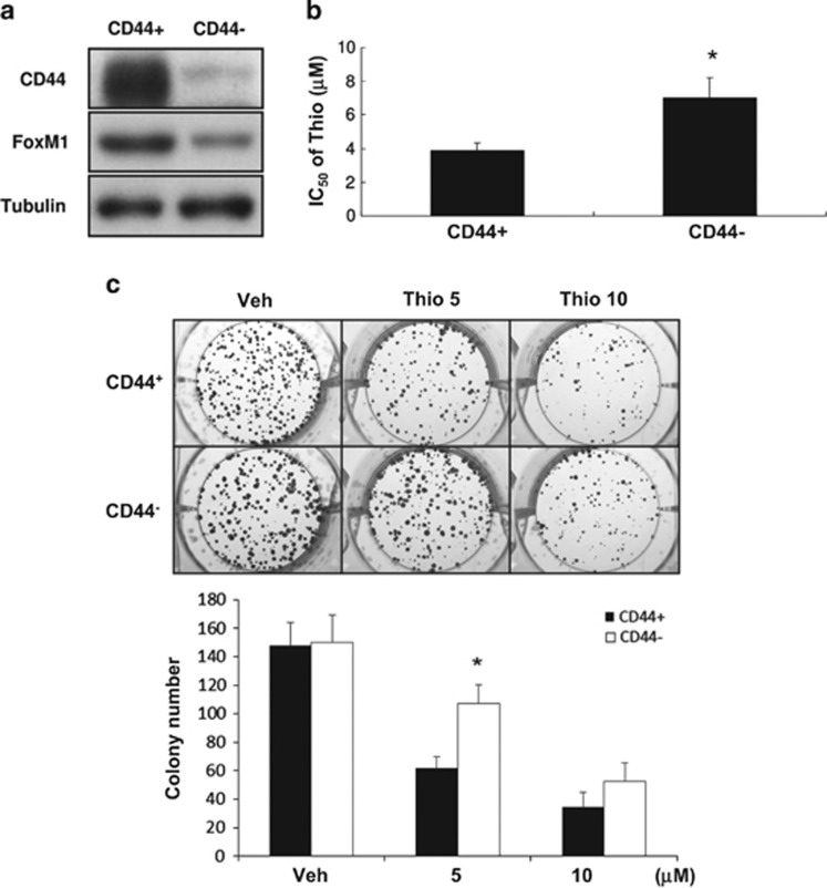
Figure 7.
Higher expression of FoxM1 is found in CD44+ HCT-15 cells, which are more sensitive to thiostrepton. (a) Protein levels of CD44 and FoxM1 in CD44+ and CD44− HCT-15 cells were analyzed by western blotting. Tubulin signal was used as a loading control. (b) The IC50 values of thiostrepton on CD44+ and CD44− HCT-15 cells were determined as described. *P<0.05 by Student's t-test. (c) A colony-formation assay was carried out by treating cells with DMSO (Veh) or 5 or 10 μM thiostrepton for 6 h before their washout of drug or control. Cells were cultured in regular media for 10 more days and colonies were stained with crystal violet. Data representing the mean±S.D. of three independent experiments were analyzed by Student's t-test (*P<0.05)

