Figure 6.
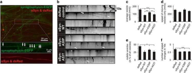
Axonal transport of synaptophysin-positive vesicles in PMN transfected with different αSyn variants. (a) Representative micrograph showing PMN transduced with AAV.synaptophysin-EGFP (expressing EGFP-tagged synaptophysin) and co-transfected with p.αSyn-WT and p.dsRed (to identify transfected neurons) (DIV 5). In the higher magnification pictures, the EGFP-positive vesicles (arrows) can be seen along the transfected axon. (b) Representative kymographs of the movements of EGFP-tagged synaptophysin along neurites transduced with the plasmids given on the left side within 10 s (y axis). Arrows point at representative moving vesicles, asterisks mark stationary vesicles. (c–f) Quantifications of synaptophysin-EGFP transport in neurites transfected with the given plasmids. Statistics: one-way ANOVA followed by Dunnett's post hoc test, *P<0.05, **P<0.005, ***P<0.0005. Error bars represent means±S.E.M. n=3 independent experiments

