Figure 2.
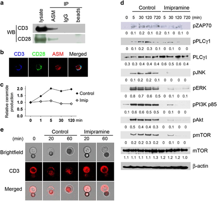
Association of ASM and CD3/CD28 in CD4+ T cells. (a) Co-immunoprecipitation of ASM with CD3 and CD28, respectively, from naive CD4+ T-cell lysates. Mouse IgG or beads only served as capture controls. (b) Colocalization of CD3/CD28 with ASM on naive CD4+ T cells was determined using confocal microscopy. (c–e) Naive CD4+ T cells were activated with anti-CD3/CD28 antibody-coated beads in the absence or presence of imipramine (20 μM) for different times, ceramide production, intracellular CD3/CD28 signal transduction, and CD3 dynamics were examined by TLC (c), western blot using specific antibodies as indicated (d), and confocal microscopy (e), respectively. The beads adjacent to cells show reflected fluorescence. Data are representative of three to five independent experiments

