Figure 4.
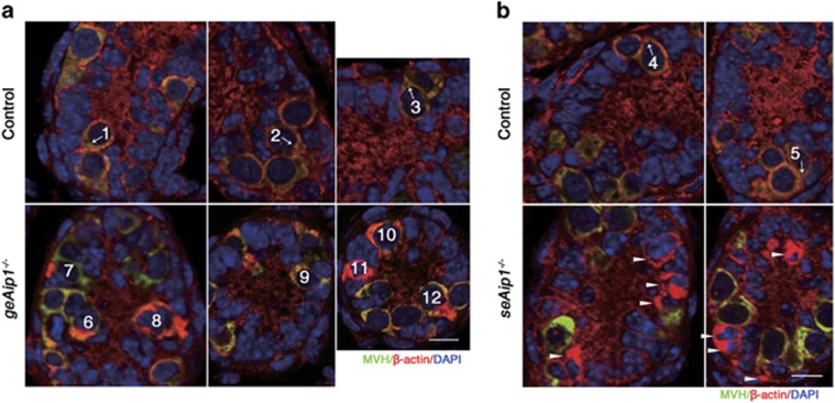
Aip1 deletion caused ectopic actin accumulations in the cell cortical regions and within lamellipodial protrusions during postnatal testis development. (a and b) Immunostaining of MVH (green) and β-actin (red) of testis tissue sections from P5 control, geAip1−/− and seAip1−/− mice. The cells numbered 1–5 in the controls are wild-type germ cells migrating toward the basement membrane. Each arrow points to the migration direction and the polarized protrusion of the numbered germ cell. The cells numbered 6–12 are Aip1-deleted germ cells containing brightly stained actin patches in cell cortical regions. Arrowheads point to Aip1-ablated Sertoli cells with ectopic actin patches. Cell nuclei were stained with DAPI (blue). Scale bars: 10 μm

