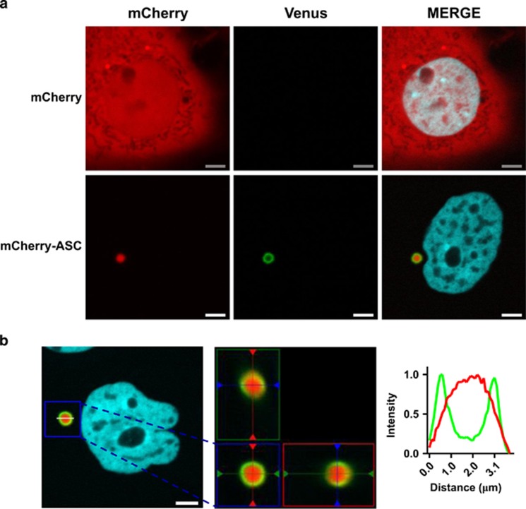
Figure 5.
Caspase-1 is dimerized on the outer surface of ASC complexes. MCF-7 cells were transiently transfected with expression plasmids encoding C1-Pro VC (40 ng), C1-Pro VN (40 ng) and mTurquoise-histone H2A-10 (blue), which localizes to the nucleus (10 ng), with mCherry (800 ng) or ASC-mCherry (800 ng) in the presence of qVD-OPH (20 μM). After 24 h, cells were assessed for caspase-1 BiFC (green) and ASC (red) colocalization. Scale bars represent 5 μm. (b) 3D reconstructions composed from 0.1 μm serial confocal images through the z plane of the cell were made. The orthogonal slice view of the boxed region (left) is shown (center). The middle panel is the xy plane, the right panel is the yz plane and top panel is the xz plane. The yz and xz planes intersect according to the cross hairs. The line scan (right) indicates the localization of C1-Pro BiFC relative to mCherry-ASC and corresponds to the line drawn on the image (left)

