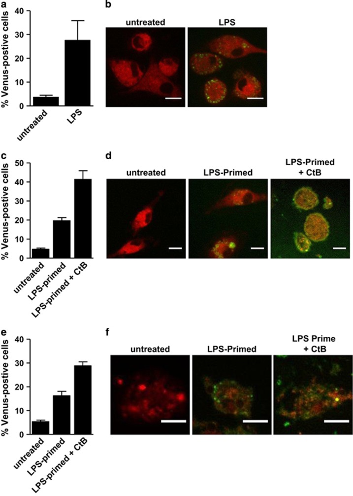
Figure 6.
Proinflammatory stimuli induce inflammatory caspase BiFC in primary murine BMDM. (a) Murine BMDM were transiently transfected with expression plasmids encoding C1-Pro VC (300 ng) and C1-Pro VN (300 ng) with dsRed as a reporter for transfection. After 24 h, cells were treated with or without ultrapure LPS (100 ng/ml) for 4 h. The percentage of transfected cells that were Venus-positive cells was determined by flow cytometry. Error bars represent the S.D. of three independent experiments. (b) Representative confocal images of cells from (a) are shown. Scale bars represent 10 μm. (c) Murine BMDM were transiently transfected with expression plasmids encoding C5-Pro VC (300 ng) and C5-Pro VN (300 ng) with dsRed as a reporter for transfection. After 24 h, cells were left untreated or primed with ultrapure LPS (100 ng/ml) for 4 h. LPS-primed cells were then treated with CtB (20 μg/ml) for 16 h. The percentage of transfected cells that were Venus-positive cells was determined by flow cytometry. Error bars represent the S.D. of three independent experiments. (d) Representative confocal images of cells from (c) are shown. Scale bars represent 10 μm. (e) Murine BMDM were transiently transfected with expression plasmids encoding C1-Pro VC (300 ng) and C5-Pro VN (300 ng) with dsRed as a reporter for transfection. Cells were treated and assessed as in (c). (f) Representative confocal images of cells from (e) are shown. Scale bars represent 50 μm

