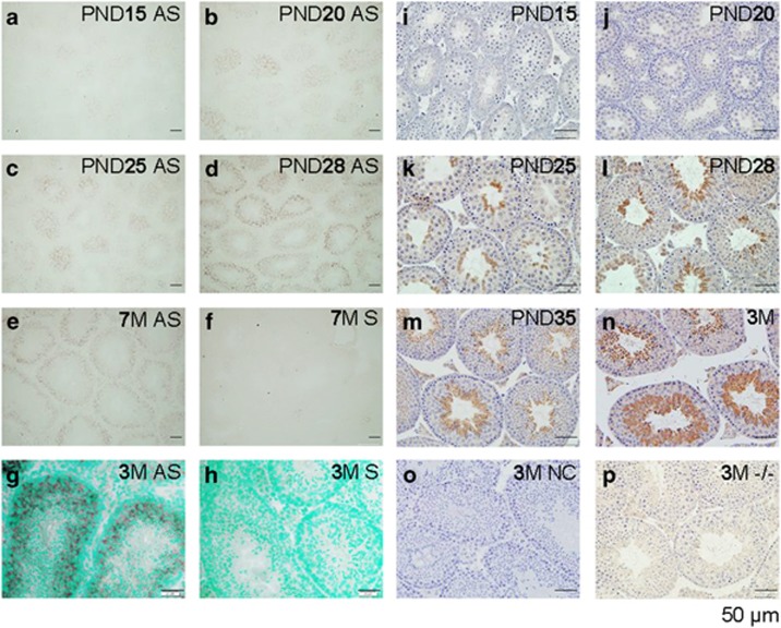
Figure 3.
Spatiotemporal localization of Bscl2 mRNA by in situ hybridization (a–h) and seipin protein by immunohistochemistry (i–p) in the testis. All sections were from Bscl2+/+ testes except p, which was from a Bscl2−/− testis. Testes from at least three mice at each time point were analyzed. Representative images were shown. (a) PND15, antisense (AS) probe. (b) PND20, antisense probe. (c) PND25, antisense probe. (d) PND28, antisense probe. (e) 7-Month-old (7M), antisense probe. (f) 7M, sense probe, negative control. (g) 3M, antisense probe. (h) 3M, sense probe. (g and h) Counterstained with methyl green. (i–p) All sections were incubated with primary anti-seipin antibody, except o, which was incubated with normal rabbit IgG. (i) PND15. (j) PND20. (k) PND25. (l) PND28. (m) PND35. (n) 3M. (o) 3M, negative control (NC). (p) 3M, Bscl2−/− (−/−), a second negative control. Scale bar, 50 μm

