Figure 3.
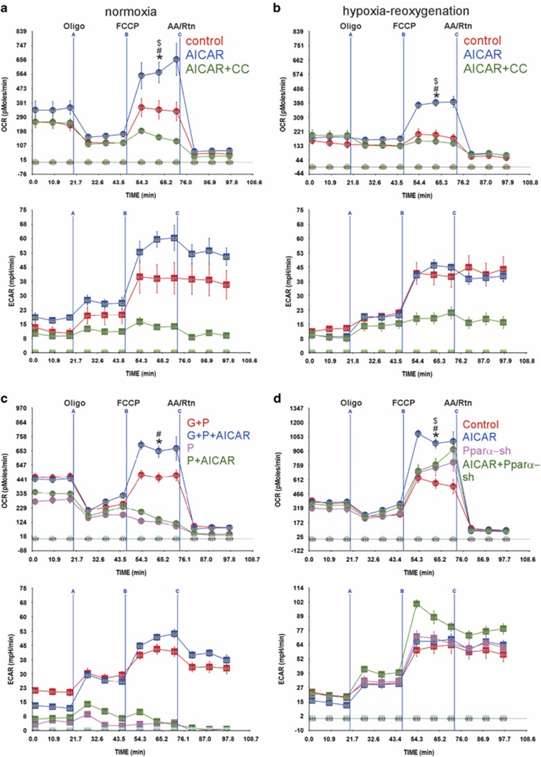
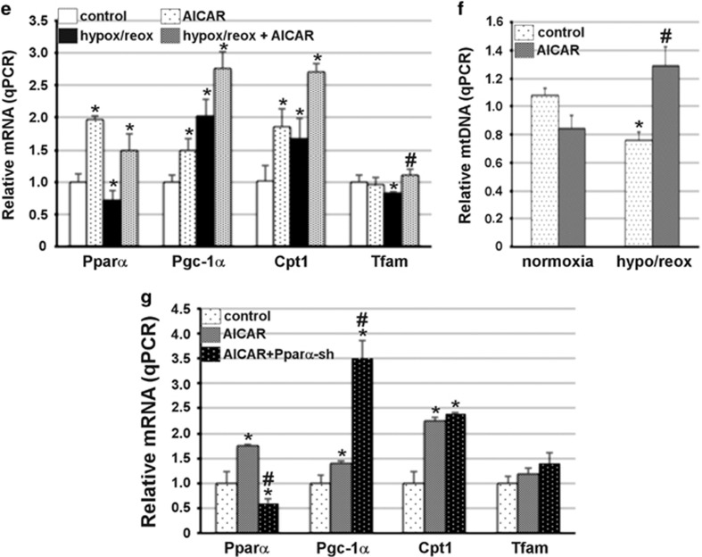
The RRC is regulated by AMPK via Pparα. (a and b) Neonatal rat cardiac myocytes were cultured in complete growth medium containing vehicle, 500 μM AICAR or 500 μM AICAR plus 5 μM compound C (CC) and either remained in normoxic conditions (atmospheric O2) (a) or were exposed to hypoxia (<1% O2), for 24 h (b). The medium was replaced with base medium containing 17.5 mM glucose plus 100 μM palmitate-BSA containing vehicle, 500 μM AICAR or 500 μM AICAR plus 5 μM CC, for 24 h. The medium was then replaced with serum-free XF medium containing 17.5 mM glucose plus 100 μM palmitate-BSA containing vehicle, 500 μM AICAR or 500 μM AICAR plus 5 μM CC, for 1 h. The mitochondrial stress test was then performed as described in Materials and Methods, n=4–6, each experiment was performed three times. Error bars represent S.E.M., *P<0.05 max OCR versus basal OCR for AICAR-treated cells, at the time point indicated; #P<0.05 max OCR for AICAR-treated versus max OCR for control, at the time point indicated; #P<0.05 max OCR for AICAR treated versus max OCR for CC treated. (c) Neonatal rat cardiac myocytes were treated with vehicle or 500 μM AICAR in the presence of 17.5 mM glucose plus 100 μM palmitate-BSA or 100 μM palmitate-BSA, for 24 h. The medium was then replaced with serum-free XF medium containing 17.5 mM glucose plus 100 μM palmitate-BSA or 100 μM palmitate-BSA containing vehicle or 500 μM AICAR, for 1 h. The mitochondrial stress test was then performed as described in Materials and Methods, n=4–6, each experiment was performed three times. Error bars represent S.E.M., *P<0.05 max OCR versus basal OCR for AICAR-treated cells in glucose+palmitate (G+P), at the time point indicated; #P<0.05 max OCR for AICAR treated versus max OCR for untreated in G+P, at the time point indicated. (d) Neonatal rat cardiac myocytes were cultured in complete growth medium treated with vehicle or 500 μM AICAR in the presence of adenoviral-control shRNA or -shRNA-Pparα- constructs, for 24 h. The medium was then replaced with serum-free XF medium containing 17.5 mM glucose plus 100 μM palmitate-BSA containing vehicle or 500 μM AICAR, for 1 h. The mitochondrial stress test was then performed as described in Materials and Methods, n=4–6, each experiment was performed two times. Error bars represent S.E.M., *P<0.05 max OCR versus basal OCR for AICAR-treated cells, at the time point indicated; #P<0.05 max OCR for AICAR treated versus max OCR for control; $P<0.05 max OCR for AICAR treated versus max OCR of AICAR- plus shRNA-Pparα-treated, at the time point indicated. (e and f) Neonatal cardiac myocytes were treated as described in (a and b), with AICAR in normoxia versus hypoxia/reoxygenation (hypox/reox), as indicated. Total mRNA (e) and DNA (f) were extracted and subjected to qPCR for the indicated genes (n=4). The results were averaged and plotted as relative values to control medium (no glucose or fatty acids). Error bars represent S.E.M., *P<0.05 versus control, #P<0.05 versus control hypox/reoxx. (g) Neonatal myocyes were treated with adenoviral shRNA-control and shRNA-Pparα, with or without 500 μM AICAR for 24 h in the presence of 17.5 mM glucose+100 μM palmitate-BSA. Total RNA was extracted and analyzed by qPCR for the indicated genes. The results were averaged and plotted as values relative to control. Error bars represent S.E.M., *P<0.05 versus control


