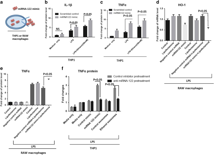
Figure 7.
Simulation experiments to confirm production of pro- inflammatory cytokines due to miRNA-122 transfer and preventing inflammatory effects of ethanol exosomes using exosome-mediated delivery of RNAi. A) To confirm that horizontal transfer of miRNA-122 is inducing pro-inflammatory phenotype in monocytes, we designed “simulation experiments” in which miRNA-122 mimic was introduced to THP1 cells and RAW macrophages via electroporation and transfection reagents, respectively. B) MiRNA-122 mimic and control mimic were introduced to the THP1 cells with electroporation (300 kV and 1500 μF), after 18 h, 10 nM LPS was added for 6 h, supernatants were collected for IL-1β ELISA analysis. The results represent three independent experiments and expressed as IL-1β protein levels fold change. C) MiRNA-122 mimic and control mimic were introduced to the cells with electroporation (300 kV and 1500 μF), after 18 h, 10 nM LPS was added for 6 h and after that supernatants were collected for TNFα ELISA analysis. The results represent three independent experiments expressed as TNFα protein level fold change. D) miRNA-122 and negative control were introduced to the RAW macrophages with Lipofectamine® RNAiMAX transfection reagent and LPS (10 nM) was added 6 h before readings. After 48 h, HO-1 mRNA levels were measured by quantitative real-time PCR. 18S was used to normalize the Ct values between the samples. E) miRNA-122 and negative control were introduced to the RAW macrophages with Lipofectamine® RNAiMAX transfection reagent and LPS (10 nM) was added 6 h before readings. After 48 h, TNFα protein levels were measured by quantitative real-time PCR. F) miRNA-122 inhibitor was loaded into THP1-derived exosomes as described in the method section. Exosomes were added to naïve THP1 cells for 12 h; after 12 h exosomes were washed off and media was replaced. Exosomes derived from ethanol-treated Huh7.5 cells were added for 8 h. TNFα ELISA were done after 24 h and 6 h before readout 10 nM LPS was added to pertinent groups. MiRNA-122 mimic was electroporated to the THP1 cells as a positive control. The results represent three independent experiments.

