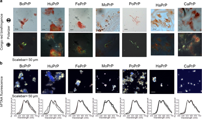
Figure 2. Microscopy of mammalian PrP aggregates stained with amyloid dyes.
(a) All PrP sequences were stained with Congo red and displayed anomalous colors i.e. birefringence under crossed polarizers. (b) All PrP sequences display amyloidotypic hFTAA fluorescence spectra (peaks at 545 nm and 585 nm indicate amyloid structure) when analyzed using hyperspectral imaging32. The spectral profile of BoPrP and HuPrP display higher spectral heterogeneity indicating more variable fibril morphology for these sequences compared to the other PrPs34.

