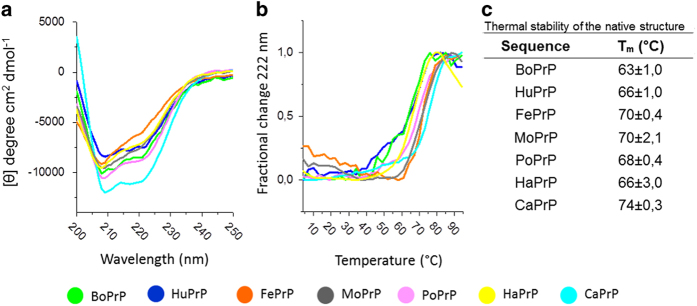
Figure 3. Circular dichroism spectroscopy of mammalian PrPs.
(a) Far UV CD spectra at 4 °C reveal high α-helical content and indicate natively folded PrPs. (b) Thermal denaturation monitored at 222 nm shows cooperative unfolding and some difference in thermal stability between the PrPs. (c) Tm values calculated according to60. Data was recorded from samples in a 1 mm cuvette with 5 μM PrP in 100 mM sodium phosphate, 50 mM NaCl, 50 mM KCl, pH 7.4.

